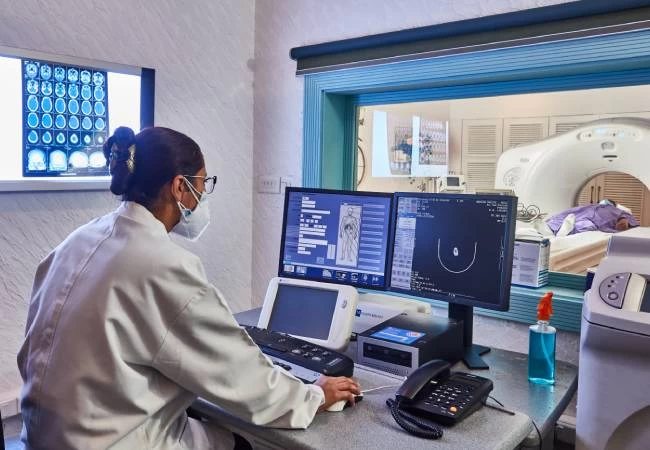
About Interventional Procedures


At Mahajan Imaging & Labs, Ultrasound and CT guided FNAC (Fine Needle Aspiration Cytology), biopsies, and drainage procedures are done routinely. It is a referral site for difficult CT guided biopsies of the chest, spine, and abdomen.
The guided biopsy uses advanced computer programs in order to create detailed images of inside the body. It is a minimally invasive procedure which is usually an outpatient procedure (OPD).
Image-guided biopsies are an OPD (Outpatient) procedure, and in most cases, the patient is able to go back home after the test. Image guidance improves diagnostic accuracy and reduces the number of attempts and consequently, the side effects of a biopsy procedure. This helps in getting a pathological diagnosis of diseases which affect deep-situated organs in the body, and many times an invasive surgical procedure may be avoided based on the results obtained.
Two to four samples of tissue are taken using a needle which is guided into an abnormality by ultrasound, CT, or mammography. These are then reviewed by the pathologist, who will provide a report to your referring Doctor. Tissue sampling uses a needle which is guided into an abnormality by ultrasound, CT, or mammography. Ultrasound is used to guide biopsies of the breast, thyroid, liver and superficial lymph nodes, and other accessible structures. CT is used to guide biopsies of the chest, abdomen, and pelvis. Some breast biopsies are guided by mammography if the abnormality is not visible on ultrasound.
This type of core needle biopsy involves ultrasound - an imaging method that uses high-frequency sound waves to produce precise images of structures within your body.
This type of biopsy uses mammograms to pinpoint the location of suspicious areas within the breast.
It is a simple, quick, and inexpensive method that is used to sample superficial masses like those found in the neck and is usually performed in the outpatient clinic. It causes minimal trauma to the patient and carries virtually no risk of complications. Masses located within the region of the head and neck, including salivary gland and thyroid gland lesions, can be readily diagnosed using this technique.
At the time of the breast biopsy procedures noted above, a tiny stainless-steel marker or clip may be placed in your breast at the biopsy site. This is done so that if your biopsy shows cancer cells or precancerous cells, your doctor or surgeon can locate the biopsy area to remove more breast tissue surgically.
Paracentesis is a procedure in which a needle or catheter is inserted into the peritoneal cavity to obtain ascitic fluid for diagnostic or therapeutic purposes. Ascitic fluid may be used to help determine the etiology of ascites as well as to evaluate for infection or the presence of cancer.
All interventional procedures at Mahajan Imaging & Labs are performed by experienced medical professionals and are generally safe. Though complications are possible but are relatively rare. There are safety protocols in place to ensure the best diagnostic outcomes and minimized risks. At Mahajan Imaging & Labs, we prioritize patient safety and adhere to the highest testing standards during every procedure.
At Mahajan Imaging & Labs, the average duration for an interventional procedure depends on the availability of the doctor/equipments for the type of scan. However, with appointment an interventional procedure scan typically takes 30 minutes.
Results for an interventional procedure typically become available within 24 working hours of the scan being completed.
Most interventional procedures are minimally painful, as any discomfort is usually managed through local sedation or anaesthesia.
The recovery time after an interventional radiology procedure depends on the patient's overall health, specific procedure, and any possible complications.
An appointment for an interventional procedure can be conveniently booked at Mahajan Imaging & Labs through our official website www.mahajanimaging.com, call our customer care number at +91 11 4118 3838 (from 7:00 A.M. to 10:30 P.M.), or chat with our WhatsApp bot at +91 88828 97661.
Usage of anaesthesia and sedation typically depends on the type of interventional procedure. Most interventional procedures are less invasive and can be performed with local anaesthesia, light general anaesthesia, or conscious sedation. It primarily depends on the patient's comfort level and procedure's complexity. At Mahajan Imaging & Labs, our experienced team will determine the most appropriate option for the patient.
Interventional procedures often require the use of small catheters and incisions that help in reducing pain, time, and infection risk. These are minimally invasive compared to the traditional surgeries as they typically require less anaesthesia and involve advanced imaging techniques.
Patients may be requested to fast for certain hours and avoid consuming certain medications prior the procedure. It becomes essential to consult with the diagnostic center or the healthcare professional in such cases.
7-B, Upper Ground Floor, Main Pusa Road, Rajinder Nagar, New Delhi - 110005
Ground Floor, Times Square Building, Sushant Lok Phase-I, Gurugram, Haryana - 122002
Fortis Hospital: Aruna Asaf Ali Marg, Pocket 1, Sector B, Vasant Kunj, New Delhi-110070
Pushpawati Singhania Research Institute (PSRI): Pocket J, Sheikh Sarai Phase II, New Delhi-110017
Fortis Escorts Hospital: Sector 5, Malviya Nagar, Jaipur, Rajasthan-302017